NIfTI Datasets
NIfTI (Neuroimaging Informatics Technology Initiative) is an open file format specifically engineered to store, manipulate, and analyze multi-dimensional (3D and 4D) neuroimaging and medical research data. Developed in the early 2000s under the sponsorship of the National Institutes of Health (NIH) as an evolution of the older ANALYZE 7.5 format, NIfTI serves as the de facto standard for neuroimaging research pipelines, scientific computing, and deep learning workflows.
NIfTI files typically have the following characteristics:
- File Structure: NIfTI data is packaged as a single consolidated file (.nii or gzip-compressed .nii.gz) containing both the header and binary voxel array.
- Coordinate Systems & Affine Transforms: A core feature of NIfTI is its rigorous spatial orientation encoding. It includes affine transformation matrices (qform and sform) that map voxel indices (i, j, k) directly to real-world continuous spatial coordinates (x, y, z) in millimeter space (standardized to a right-handed RAS or LPS system).
- Multi-Dimensional Support: While the first three dimensions describe 3D space (x, y, z), the 4th dimension typically represents time points (t), and higher dimensions can hold vector-valued or multi-component data.
NIfTI is primarily utilized across brain imaging and biomedical research modalities:
- Structural MRI (3D): High-resolution anatomical scans.
- Functional MRI (fMRI, 4D): Time-series BOLD (Blood-Oxygen-Level-Dependent) scans capturing brain activation over time.
- Diffusion MRI (dMRI / DTI, 4D): Scans measuring water diffusion across multiple gradient directions to reconstruct neural tractography.
- Volumetric CT & PET: 3D attenuation or metabolic emission maps used in quantitative research.
- Segmentation Masks & Annotations: Binary or multi-class label volumes representing anatomical structures or lesions.
DICOM Studio supports the visualization of images in NIfTI Format in 2D and 3D. Conversion of DICOM Images to a single NIfTI file is also supported. The only difference in DICOM Studio when working with NIfTI Datasets compared to DICOM Studies, is Study Management. This is not supported.
Import NIfTI Dataset
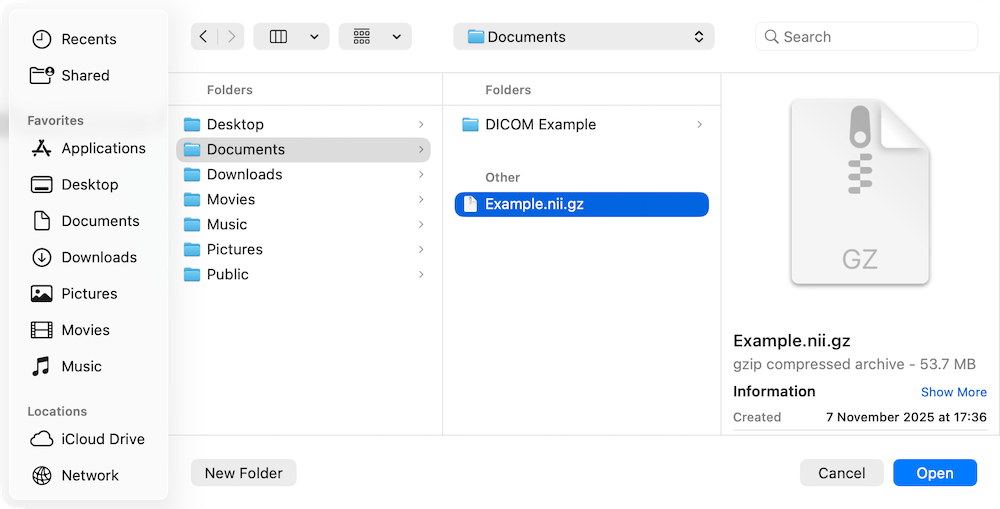
From the menubar, open the File menu and select Open NIfTI File. Your device's file explorer will open. Navigate to the location where you want to save the .nii or nii.gz file. Select the file and click Open.
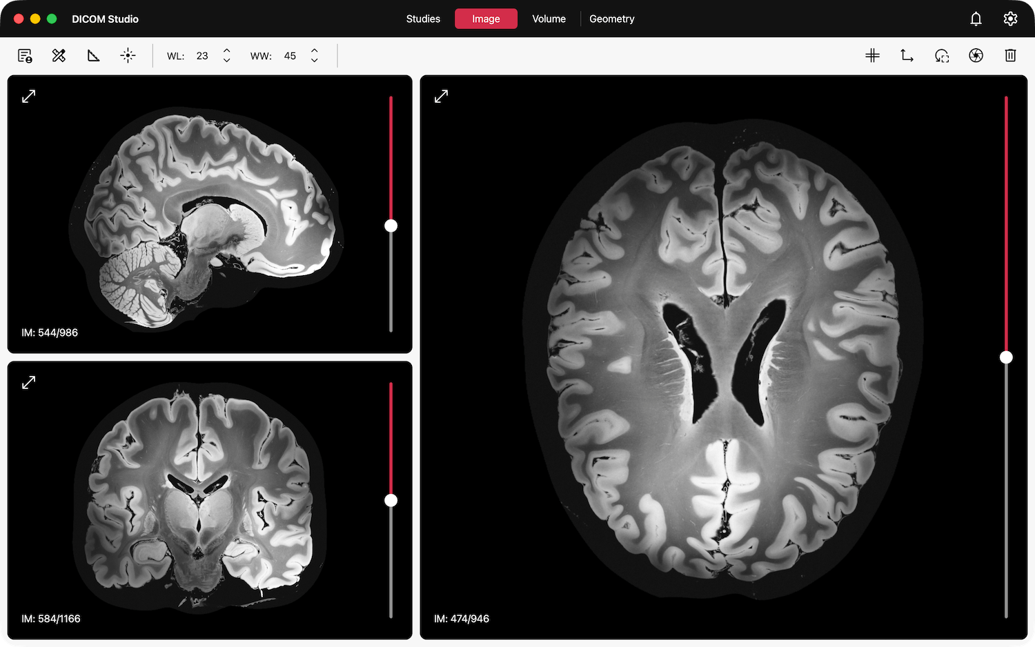
Slices in all 3 orientations will be loaded onto the three views in the Image tab. Images in the original orientation will be loaded onto the primary view on the right. Slices in the other 2 orientations are reconstructed, and loaded onto the remaining 2 views on the left. Learn more on how to interact with the image views, and analyze your images, in the 2D Imaging section.
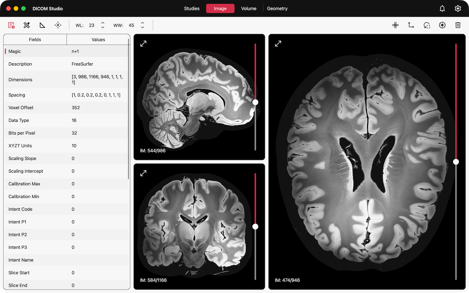
The metadata from the NIfTI Header will be populated on the table to the left, accessible by clicking the Show DICOM Tags / NIfTI Headers button on the toolbar with the icon below.
All 2D & 3D features described in the 2D Imaging, Volume Rendering and Geometric Modelling sections will now be available.
Convert a DICOM Series to a NIfTI File
The steps to follow are outlined in detail in the Share Studies Section, here. What we will cover here are the settings available to this conversion.
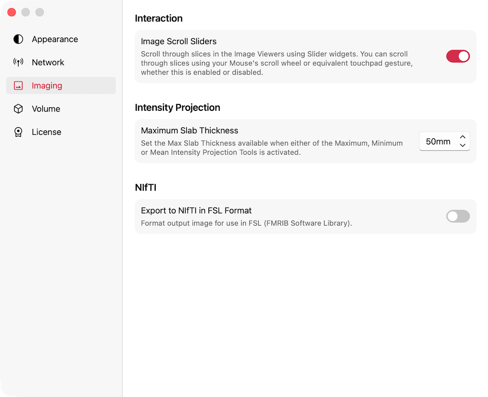
Under Imaging -> NIFTI , there is a Export to NIfTI in FSL Format switch. When this is off, the final image after conversion will have the same orientation as the source DICOM Images. When this is on, the conversion process will format an axial image for use in FSL.
FSL (FMRIB Software Library) is a comprehensive library of analysis tools for FMRI, MRI and DTI brain imaging data, developed by the Oxford Centre for Functional MRI of the Brain (FMRIB).