What's New in DICOM Studio Desktop v2.3
We are thrilled to announce the release of DICOM Studio version 2.3 on Desktop. This major update brings a transformative redesign to our user interface, alongside a powerful suite of new features engineered to elevate your medical imaging experience. Whether you are conducting complex anatomical analyses, managing vast study databases, or integrating with enterprise servers, version 2.3 is built to make your workflow more intuitive, efficient, and precise.
Here is a closer look at what's new.
A Radically Redesigned, Workspace-Driven UI
To provide a more focused and uncluttered environment, we have fundamentally restructured DICOM Studio into four core functional areas: Studies, Image, Volume, and Geometry. Instead of navigating through complex menus, each tab now acts as its own dedicated workspace, equipping you with a specialized set of tools perfectly tailored to the task at hand. This modular approach minimizes cognitive load and allows you to seamlessly transition between study management, 2D analysis, 3D rendering, and geometric measurements.
The All-New Studies Tab: Your Unified Management Hub
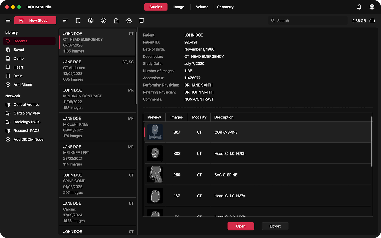
Say goodbye to disorganized data. The new Studies Tab serves as a comprehensive, unified hub designed to keep your imaging data meticulously organized. From this centralized workspace, you can effortlessly sort, save, anonymize, edit, and export your DICOM studies. It is built from the ground up to streamline administrative tasks and ensure that your data is always exactly where you need it, when you need it.
Seamless DIMSE and DICOMWeb Integration
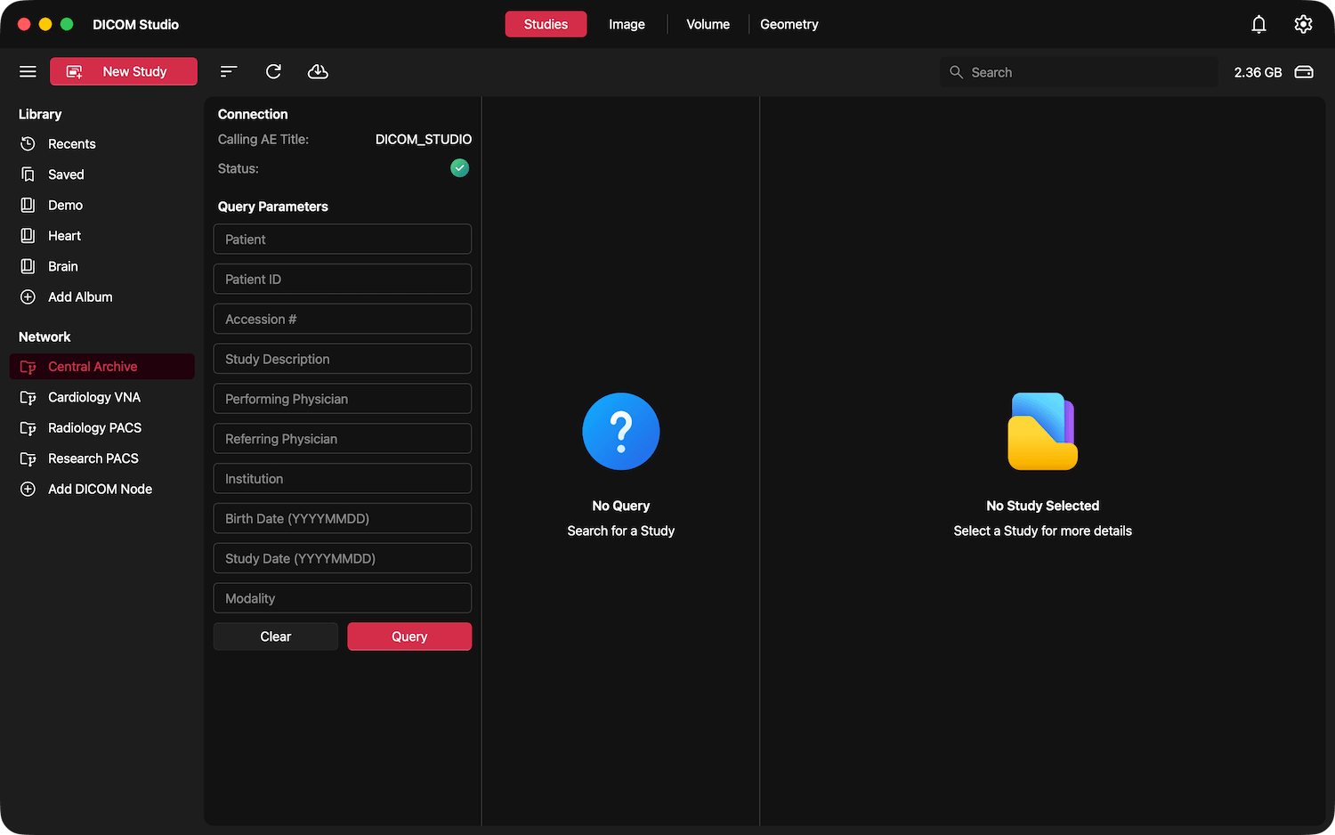
Connectivity is crucial in modern medical environments. With version 2.3, we have introduced robust DIMSE and DICOMWeb integration. You can now seamlessly query and retrieve studies directly from your networked servers without ever leaving the application. Furthermore, the new update allows you to securely upload and push imported studies back to your servers, bridging the gap between local analysis and enterprise-wide image archives.
Immediate Context with Multi-Planar Views
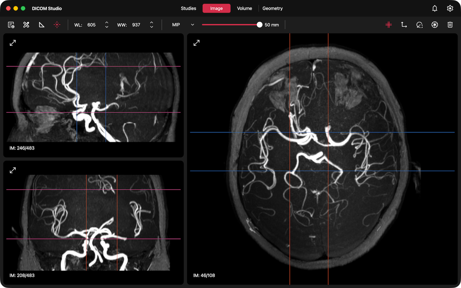
To provide instant anatomical context the moment you open a study, newly loaded images are now displayed in all three primary orientations by default. Within the same window, you will immediately see three separate, synchronized views: Axial, Sagittal, and Coronal. DICOM Studio automatically generates the two additional orthogonal planes from your original image using Multi-Planar Reconstruction (MPR), providing immediate 3D spatial context from the very first click.
Advanced Intensity Projections (MIP, MinIP, and MeanIP)
Enhance your diagnostic clarity and image analysis capabilities with our new projection tools, located within the Image Tab. You can now easily generate Maximum Intensity Projections (MIP) to visualize hyper-dense structures like contrast-enhanced vessels or bone, Minimum Intensity Projections (MinIP) for optimal assessment of the biliary tree or airways, and Average (Mean) Intensity Projections for clearer tissue differentiation.
Pinpoint Precision with Cross-Reference Lines
Spatial orientation is critical when navigating complex multi-planar reconstructions (MPR). Our newly introduced cross-reference lines dynamically track structures and anomalies across different viewing planes. By providing immediate, real-time spatial correlation between the axial, coronal, and sagittal planes, this feature allows practitioners to confidently localize anatomical features and pathology.
Native NIfTI File Support
Recognizing the diverse formats used in medical research and neuroimaging, DICOM Studio v2.3 now natively supports the import and visualization of NIfTI images (.nii, .nii.gz). Not only can you seamlessly view these files alongside your standard images, but our new export engine also allows you to convert any standard DICOM series directly into NIfTI format from the Studies Tab.
Volume Lasso Selection Crop Tool
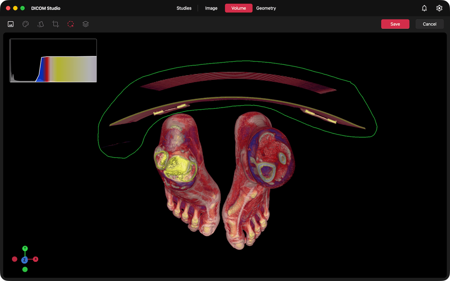
While our existing box crop widget provides excellent utility for cropping 3D volume objects along the x, y, or z axes, we wanted to offer even greater flexibility for isolating anatomical structures. With the new Lasso Crop tool, you are no longer constrained by rigid geometric planes. You can now simply draw freehand over any part of the volume you wish to cut, and that exact section will be seamlessly cropped in 3D. This intuitive approach makes it incredibly easy to remove hard-to-reach areas of the volume and focus purely on the anatomy that matters most.
A More Robust DICOM Reader
Beneath the hood, our core DICOM reading engine has undergone a massive overhaul. It is now significantly more resilient, and capable of handling a significantly wider array of image formats and edge cases. Whether you are dealing with non-standard encodings or massive, complex multi-frame studies, the enhanced DICOM Reader ensures that your files load reliably and render flawlessly.
Experience the Next Generation of DICOM Studio
DICOM Studio v2.3 represents our ongoing commitment to empower medical practitioners with world-class, accessible, and affordable medical imaging software. We invite you to download the latest update today and experience a faster, more organized, and deeply capable imaging platform.
— The DICOM Studio Team
Work seamlessly across all your devices.
Whether in the radiology suite, at the point of care, or on the go, DICOM Studio delivers a consistent, high-performance experience.